Subarea 1: Stem Cell Aging
The individual research groups within Subarea 1 investigate the causes and consequences of stem cell aging. The research work spans from basic model organisms over genetic mouse models up to humanized mouse models engrafted with human stem cells.
According to the FLI, with the closure of two groups since 2016 the representation of invertebrate models of stem cell research was reduced in Subarea 1. The institute presumes that the recruitment of new groups should fill this gap.
The research is defined by four focus areas:
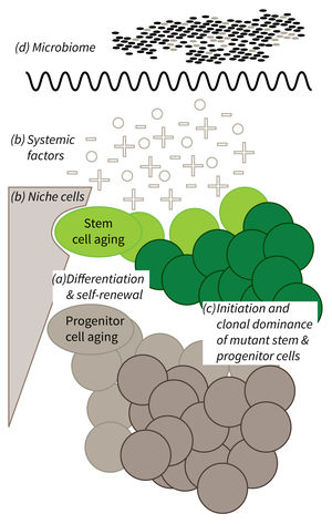
- Cell-intrinsic mechanisms limiting the function of aging stem and progenitor cells,
- Aging-associated alterations of stem cell niches and the systemic environment,
- Mechanisms of clonal selection and epigenetic drifts in stem cell aging, and
- Microbiota- and metabolism-induced impairments in stem cell function during aging (in context of the new focus area Microbiota and Aging currently being built up within Subarea 2).
Research focus of Subarea 1.
a) It is currently not well understood what mechanisms impair cellular functions in aging. b) The relative contribution of niche cells and systemic acting factors on stem cell aging have yet to be determined in different tissues. c) Clonal expansion of mutant cells associates with disease development in aging humans. Mechanistically, the process remains poorly understood. Changes in color intensity depict clonal dominance originating from stem (green) or progenitor cells (gray). d) Emerging evidences indicate that aging associated alter ations in microbiota influence stem cell function and vice versa.
Publications
(since 2016)
2016
- Metabolic regulation of stem cell function in tissue homeostasis and organismal ageing.
Chandel NS, Jasper H, Ho TT, Passegué E
Nat Cell Biol 2016, 18(8), 823-32 - You Are What You Eat: Linking High-Fat Diet to Stem Cell Dysfunction and Tumorigenesis.
Haller S, Jasper H
Cell Stem Cell 2016, 18(5), 564-6 - Gene Dosage Reductions of Trf1 and/or Tin2 Induce Telomere DNA Damage and Lymphoma Formation in Aging Mice.
Hartmann K, Illing A, Leithäuser F, Baisantry A, Quintanilla-Fend L, Rudolph KL
Leukemia 2016, 30(3), 749-53 - Wnt/β-catenin signaling via Axin2 is required for myogenesis and, together with YAP/Taz and Tead1, active in IIa/IIx muscle fibers.
Huraskin D, Eiber N, Reichel M, Zidek LM, Kravic B, Bernkopf D, von Maltzahn J, Behrens J, Hashemolhosseini S
Development 2016, 143(17), 3128-42 - Telomere shortening leads to earlier age of onset in ALS mice.
Linkus B, Wiesner D, Meßner M, Karabatsiakis A, Scheffold A, Rudolph KL, Thal DR, Weishaupt JH, Ludolph AC, Danzer KM
Aging (Albany NY) 2016, 8(2), 382-93 - Intestinal IRE1 Is Required for Increased Triglyceride Metabolism and Longer Lifespan under Dietary Restriction.
Luis NM, Wang L, Ortega M, Deng H, Katewa SD, Li PWL, Karpac J, Jasper** H, Kapahi** P
Cell Rep 2016, 17(5), 1207-16 ** co-corresponding authors - Loss of fibronectin from the aged stem cell niche affects the regenerative capacity of skeletal muscle in mice.
Lukjanenko L, Jung MJ, Hegde N, Perruisseau-Carrier C, Migliavacca E, Rozo M, Karaz S, Jacot G, Schmidt M, Li L, Metairon S, Raymond F, Lee U, Sizzano F, Wilson DH, Dumont NA, Palini A, Fässler R, Steiner P, Descombes P, Rudnicki MA, Fan CM, von Maltzahn J, Feige JN, Bentzinger CF
Nat Med 2016, 22(8), 897-905 - Kinomics Screening Identifies Aberrant Phosphorylation of CDC25C in FLT3-ITD-positive AML.
Perner F, Schnöder TM, Fischer T, Heidel FH
Anticancer Res 2016, 36(12), 6249-58 - p21 shapes cancer evolution.
Romanov VS, Rudolph KL
Nat Cell Biol 2016, 18(7), 722-4 - Telomere shortening leads to an acceleration of synucleinopathy and impaired microglia response in a genetic mouse model.
Scheffold A, Holtman IR, Dieni S, Brouwer N, Katz SF, Jebaraj BMC, Kahle PJ, Hengerer B, Lechel A, Stilgenbauer S, Boddeke EWGM, Eggen BJL, Rudolph KL, Biber K
Acta Neuropathol Commun 2016, 4(1), 87


