Core Facilites and Core Services
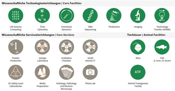
At the beginning of 2016, a “core” structure was put into effect that organized facility and service units as independent organizational entities from FLI’s research groups. A number of technology platforms (e.g. sequencing, mass spectrometry) grew out of individual methodological requirements for single research groups in the last years but developed into semiautonomous substructures. As consequence of re-focused research activities and the concomitant advent of new research groups at FLI, those units increasingly had to serve many FLI groups and collaborative research efforts in the Jena research area.
To accommodate this development and to increase efficiency as well as transparency for users, facility personnel and for administrative processes, it came natural to re-organize such activities into independent units as “FLI Core Facilities and Services” and to phase out infrastructures considered non-essential for FLI’s research focus (X-ray crystallography and NMR spectroscopy).
FLI’s Core Facilities (CF) are managed by a CF Manager and are each scientifically guided in their activities and development by an FLI Group Leader, as Scientific Supervisor. The animal facilities comprising fish, mouse and transgenesis are run separately, as they involve a more complex organizational structure. Basic Core Services (CS) are directly led by the Head of Core (HC), who in turn is supported by individual CS Managers.
All facilities and services, including animal facilities, have a valuable contribution to FLI’s research articles; e.g. from 2016–2018, to 54% of all peer reviewed research publications.
Overview Core Facilities and Core Services at FLI.
Publications
(since 2016)
2022
- Tight association of autophagy and cell cycle in leukemia cells.
Gschwind* A, Marx* C, Just MD, Severin P, Behring H, Marx-Blümel L, Becker S, Rothenburger L, Förster M, Beck JF, Sonnemann J
Cell Mol Biol Lett 2022, 27(1), 32 * equal contribution - Taz protects hematopoietic stem cells from an aging-dependent decrease in PU.1 activity.
Kim* KM, Mura-Meszaros* A, Tollot* M, Krishnan MS, Gründl M, Neubert L, Groth M, Rodriguez-Fraticelli A, Svendsen AF, Campaner S, Andreas N, Kamradt T, Hoffmann S, Camargo FD, Heidel FH, Bystrykh LV, de Haan G, von Eyss B
Nat Commun 2022, 13(1), 5187 * equal contribution - NMR of intrinsically disordered proteins: A note on the application of 15 N-13 Cα het-TOCSY mixing for 13 Cα magnetisation transfers.
Kumar A, Wiedemann C, Bellstedt P, Ramachandran R, Ohlenschläger O
J Magn Reson 2022, 337, 107166 - High-yield production of coenzyme F420 in Escherichia coli by fluorescence-based screening of multi-dimensional gene expression space.
Last D, Hasan M, Rothenburger L, Braga D, Lackner G
Metab Eng 2022, 73, 158-67 - Acetylation-Specific Interference by Anti-Histone H3K9ac Intrabody Results in Precise Modulation of Gene Expression.
Lisi S, Trovato M, Vitaloni O, Fantini M, Chirichella M, Tognini P, Cornuti S, Costa M, Groth M, Cattaneo A
Int J Mol Sci 2022, 23(16), 8892 - Telomerase deficiency reflects age-associated changes in CD4+ T cells.
Matthe DM, Thoma OM, Sperka T, Neurath MF, Waldner MJ
Immun Ageing 2022, 19(1), 16 - RNA-seq analysis of brain aging in wild specimens of short-lived turquoise killifish. Commonalities and differences with aging under laboratory conditions.
Mazzetto M, Caterino C, Groth M, Ferrari E, Reichard M, Baumgart M, Cellerino A
Mol Biol Evol 2022, 39(11), msac219 - Sendungsregister
Mohnheim F, Weinzierl I, Hoppe B, Bösger J
2022, URL https://github.com/mohnbroet - Aging Activates the Immune System and Alters the Regenerative Capacity in the Zebrafish Heart.
Reuter H, Perner B, Wahl F, Rohde L, Koch P, Groth M, Buder K, Englert C
Cells 2022, 11(3), 345. doi: 10.3390/cells11030345. - Rhodopsins build up the birefringent bodies of the dinoflagellate Oxyrrhis marina.
Rhiel E, Hoischen C, Westermann M
Protoplasma 2022, 259(4), 1047-60


