Core Facilites and Core Services
At the beginning of 2016, a “core” structure was put into effect that organized facility and service units as independent organizational entities from FLI’s research groups. A number of technology platforms (e.g. sequencing, mass spectrometry) grew out of individual methodological requirements for single research groups in the last years but developed into semiautonomous substructures. As consequence of re-focused research activities and the concomitant advent of new research groups at FLI, those units increasingly had to serve many FLI groups and collaborative research efforts in the Jena research area.
To accommodate this development and to increase efficiency as well as transparency for users, facility personnel and for administrative processes, it came natural to re-organize such activities into independent units as “FLI Core Facilities and Services” and to phase out infrastructures considered non-essential for FLI’s research focus (X-ray crystallography and NMR spectroscopy).
FLI’s Core Facilities (CF) are managed by a CF Manager and are each scientifically guided in their activities and development by an FLI Group Leader, as Scientific Supervisor. The animal facilities comprising fish, mouse and transgenesis are run separately, as they involve a more complex organizational structure. Basic Core Services (CS) are directly led by the Head of Core (HC), who in turn is supported by individual CS Managers.
All facilities and services, including animal facilities, have a valuable contribution to FLI’s research articles; e.g. from 2016–2018, to 54% of all peer reviewed research publications.
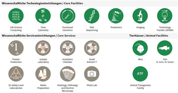
Overview Core Facilities and Core Services at FLI.
Publications
(since 2016)
2019
- Metastatic-niche labelling reveals parenchymal cells with stem features.
Ombrato L, Nolan E, Kurelac I, Mavousian A, Bridgeman VL, Heinze I, Chakravarty P, Horswell S, Gonzalez-Gualda E, Matacchione G, Weston A, Kirkpatrick J, Husain E, Speirs V, Collinson L, Ori A, Lee JH, Malanchi I
Nature 2019, 572(7771), 603-8 - Characterization under quasi-native conditions of the capsanthin/capsorubin synthase from Capsicum annuum L.
Piano D, Cocco E, Guadalupi G, Kalaji HM, Kirkpatrick J, Farci D
Plant Physiol Biochem 2019, 143, 165-75 - Analysis of the coding sequences of clownfish reveals molecular convergence in the evolution of lifespan.
Sahm A, Almaida-Pagán P, Bens M, Mutalipassi M, Lucas-Sánchez A, de Costa Ruiz J, Görlach M, Cellerino A
BMC Evol Biol 2019, 19(1), 89 - Structural insights into heme binding to IL-36α proinflammatory cytokine.
Wißbrock* A, Goradia* NB, Kumar A, Paul George AA, Kühl T, Bellstedt P, Ramachandran R, Hoffmann P, Galler K, Popp J, Neugebauer U, Hampel K, Zimmermann B, Adam S, Wiendl M, Krönke G, Hamza I, Heinemann SH, Frey S, Hueber AJ, Ohlenschläger** O, Imhof** D
Sci Rep 2019, 9(1), 16893 * equal contribution, ** co-corresponding authors
2018
- Cell-based RNAi screening and high-content analysis in primary calvarian osteoblasts applied to identification of osteoblast differentiation regulators.
Ahmad M, Kroll T, Jakob J, Rauch A, Ploubidou A, Tuckermann J
Sci Rep 2018, 8(1), 14045 - A new RelB-dependent CD117+ CD172a+ murine dendritic cell subset preferentially induces Th2 differentiation and supports airway hyperresponses in vivo.
Andreas N, Riemann M, Castro CN, Groth M, Koliesnik I, Engelmann C, Sparwasser T, Kamradt T, Haenold R, Weih F
Eur J Immunol 2018, 48(6), 923-36 - Transcriptomic alterations during ageing reflect the shift from cancer to degenerative diseases in the elderly.
Aramillo Irizar P, Schäuble S, Esser D, Groth M, Frahm C, Priebe S, Baumgart M, Hartmann N, Marthandan S, Menzel U, Müller J, Schmidt S, Ast V, Caliebe A, König R, Krawczak M, Ristow M, Schuster S, Cellerino A, Diekmann S, Englert C, Hemmerich P, Sühnel J, Guthke R, Witte OW, Platzer M, Ruppin E, Kaleta C
Nat Commun 2018, 9(1), 327 - Naked mole-rat transcriptome signatures of socially suppressed sexual maturation and links of reproduction to aging.
Bens* M, Szafranski* K, Holtze S, Sahm A, Groth M, Kestler HA, Hildebrandt** TB, Platzer** M
BMC Biol 2018, 16(1), 77 * equal contribution, ** co-senior authors - Spatial tissue proteomics quantifies inter- and intra-tumor heterogeneity in hepatocellular carcinoma.
Buczak* K, Ori* A, Kirkpatrick JM, Holzer K, Dauch D, Roessler S, Endris V, Lasitschka F, Parca L, Schmidt A, Zender L, Schirmacher P, Krijgsveld J, Singer** S, Beck** M
Mol Cell Proteomics 2018, 17(4), 810-25 * equal contribution, ** co-corresponding authors - On the S-layer of Thermus thermophilus and the assembling of its main protein SlpA.
Farci D, Farci SF, Esposito F, Tramontano E, Kirkpatrick J, Piano D
Biochim Biophys Acta 2018, 1860(8), 1554-62


